-
-
2/614 A, Philips Street, ECR,
Neelankarai,Chennai-600115

Scans During Pregnancy: Complete Guide to Ultrasounds, Anomaly & 3D/4D Scans
Pregnancy scans are an important part of antenatal care because they allow healthcare professionals to assess the pregnancy at different stages and look at aspects of fetal development, growth, the placenta and other pregnancy-related findings. However, there is no single scan schedule that is appropriate for every pregnancy. The timing and number of ultrasounds can vary depending on the stage of pregnancy, previous pregnancy history, maternal health, fetal findings and the recommendation of the obstetrician.
If you are wondering when you should have your first ultrasound, what the 12th-week scan checks, when the anomaly or anatomy scan is performed, or when 3D and 4D scans may be useful, understanding the purpose of each scan can make antenatal care less confusing.
At Healtree Multi-Speciality Centre & Hospital in Neelankarai, ECR, Chennai, Obstetrics & Gynaecology care includes routine antenatal and high-risk pregnancy management. The hospital's listed specialists include Dr. Suganya R, M.B.B.S., D.N.B. (OBG), and Dr. Hemalatha, M.B.B.S., M.D. (OBG).

What Are Scans During Pregnancy?
Pregnancy scans, also called ultrasound examinations or sonograms, use sound waves to create images of the developing pregnancy. Depending on the stage of pregnancy and the reason for the examination, an ultrasound may help assess fetal development, gestational age, fetal number, placental position, growth and other pregnancy-related findings.
Unlike X-rays, ultrasound imaging uses sound waves rather than ionizing radiation. Ultrasound is widely used during pregnancy, but the scan that is appropriate for one person may not be the same as the scan recommended for another.
The purpose of an ultrasound is not simply to produce a picture of the baby. A clinically indicated scan provides information that can help the obstetrician assess how the pregnancy is progressing and determine whether additional evaluation or monitoring may be appropriate.
When Should You Have Your First Ultrasound During Pregnancy?
The timing of the first ultrasound during pregnancy depends on the pregnancy and the reason for the scan.
An early scan may be recommended when a healthcare professional needs to assess aspects such as the location of the pregnancy, gestational age, fetal development or viability. Later in the first trimester, ultrasound can also form part of first-trimester screening.
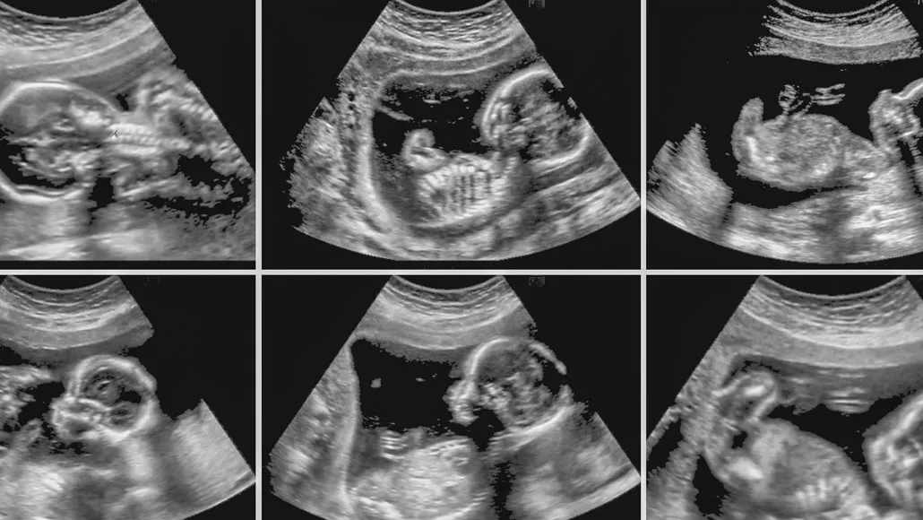
There is therefore an important distinction between:
These are not necessarily interchangeable terms.
What Is Checked During the First Pregnancy Ultrasound?
Depending on the stage and clinical reason for the scan, the ultrasound may help assess:
The 12-week or dating scan is commonly performed around the end of the first trimester in many antenatal pathways. For example, NHS guidance describes a dating scan around 10–14 weeks and notes that it can assess gestational age, fetal development and the number of babies.
Your obstetrician may recommend a different timing depending on your individual circumstances.
What Is the 12th Week Scan During Pregnancy?
The 12th week scan during pregnancy generally refers to an ultrasound performed around the first-trimester period, often as part of dating and early pregnancy assessment.
It can help establish or refine gestational age and assess fetal development. When combined with appropriate screening, an ultrasound measurement of the fluid at the back of the baby's neck, known as nuchal translucency (NT), may also be performed.
The NT measurement can form part of a screening pathway for certain chromosomal conditions. It is important to understand that screening is not the same as diagnosis. If screening indicates an increased chance of a condition, your healthcare team may discuss further testing.
What Does a 12-Week Scan Check?
Depending on the examination and clinical context, the scan may assess:
A detailed first-trimester examination is a more specialized assessment and may be indication-driven rather than something every pregnancy automatically requires. AIUM practice parameters distinguish standard obstetric ultrasound from detailed first-trimester examinations and note that examinations may vary according to the clinical situation.
What Is an Anatomy Scan During Pregnancy?
An anatomy scan during pregnancy is a detailed ultrasound examination used to assess fetal anatomy during the second trimester.
It is also commonly called an anomaly scan, mid-pregnancy scan or 20-week scan.
If you have searched for “analogy scan during pregnancy,” the term you are most likely looking for is “anomaly scan.”
The anatomy/anomaly scan is different from a basic early pregnancy scan because the fetus is further developed, allowing the healthcare professional to assess a wider range of anatomical structures.
Is an Anatomy Scan the Same as an Anomaly Scan?
In everyday antenatal care, anatomy scan and anomaly scan are often used to describe the detailed second-trimester ultrasound examination.
The terminology can vary between hospitals and healthcare systems, but the purpose is broadly to assess fetal anatomy and look for certain structural abnormalities that may be detectable at that stage.
The NHS describes the 20-week screening scan as an anomaly scan and states that it is usually performed between 18 and 21 weeks.
What Is the Best Time for an Anomaly Scan During Pregnancy?
The detailed anomaly scan is commonly performed during the second trimester, often around 18–22 weeks of pregnancy, with the exact timing determined by the obstetrician or ultrasound service.
The purpose is to examine fetal anatomy and identify certain structural conditions that may be visible at this stage. The scan may also assess fetal growth, placenta and other pregnancy-related findings. AIUM maintains specific practice parameters for detailed second- and third-trimester diagnostic obstetric ultrasound examinations.
What Does the Anomaly Scan Check?
Depending on the examination, the healthcare professional may assess structures including:
- Brain and head
- Face
- Spine
- Heart
- Chest
- Abdomen
- Kidneys and urinary system
- Limbs and bones
- Placenta
- Amniotic fluid
- Fetal growth and measurements
The exact examination depends on the clinical protocol and the circumstances of the pregnancy.
The anomaly scan can identify some structural abnormalities, but no ultrasound can detect every possible fetal or genetic condition. A scan that appears reassuring does not guarantee that a baby has no health condition.
For this reason, ultrasound findings need to be interpreted together with the mother's medical history, screening results and other clinical information.
What Is the 6th Month Scan During Pregnancy?
The phrase “6th month scan during pregnancy” is commonly used by patients to describe an ultrasound performed around the later part of the second trimester.
However, there is no universal rule that every pregnant woman must have one specific scan simply because she has reached the sixth month.
The scan schedule depends on what has already been assessed and whether the obstetrician identifies a reason for additional monitoring.
Depending on the pregnancy, an ultrasound during this period may be used to assess:
- Fetal growth
- Fetal measurements
- Placental position
- Amniotic fluid
- Fetal position
- Previously identified findings
- Areas requiring follow-up
If the earlier anatomy scan was incomplete because of fetal position or other technical limitations, a repeat examination may also be recommended.
What Scans Are Done During the Third Trimester?
Third-trimester ultrasound is not automatically identical for every pregnancy.
An obstetrician may recommend additional ultrasound examinations when there is a clinical reason to monitor fetal growth, amniotic fluid, placental findings, fetal position or another aspect of the pregnancy.
Growth Scan During Pregnancy
A growth scan can be used to assess fetal size and growth measurements.
Depending on the examination, measurements can include:
- Head measurements
- Allergy treatment
- Abdominal circumference
- Femur length
- Estimated fetal weight
- Amniotic fluid assessment
- Placental assessment
AIUM's obstetric ultrasound parameters include fetal biometry such as biparietal diameter, head circumference, abdominal circumference and femur length, along with estimated fetal weight in standard examinations.
A growth scan does not simply answer whether the baby is "big" or "small." The obstetrician considers the measurements in relation to gestational age and the overall clinical picture.
What Is a Doppler Scan During Pregnancy?
A Doppler ultrasound uses ultrasound technology to assess blood flow in selected blood vessels.
It may be recommended in pregnancies where the obstetrician needs additional information about fetal or placental circulation.
Doppler assessment is therefore not necessarily a routine scan for every pregnancy. The decision depends on the clinical circumstances.
If your doctor recommends Doppler evaluation, ask what specific blood flow is being assessed and why it is relevant to your pregnancy.
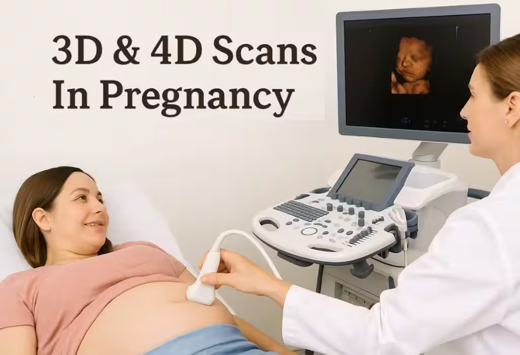
What Is the Best Time for a 3D Scan During Pregnancy?
There is no single best week for every 3D scan, because image quality depends on factors such as fetal position, amniotic fluid, gestational age and the purpose of the examination.
A 3D ultrasound creates a three-dimensional image from ultrasound data, while conventional 2D ultrasound remains central to routine diagnostic assessment.
3D imaging can provide additional visual information, but a 3D image does not automatically mean that the scan is medically more useful than a standard ultrasound.
The appropriate timing should therefore depend on the clinical purpose and the recommendation of the healthcare professional.
What Is the Best Time for a 4D Scan During Pregnancy?
A 4D ultrasound adds movement or real-time imaging to three-dimensional ultrasound.
Patients sometimes seek 4D scans to obtain more detailed images of the baby's face or movements. However, the quality of the images can vary significantly depending on fetal position, gestational age, amniotic fluid and other factors.
More importantly, 3D and 4D imaging should not replace clinically indicated diagnostic ultrasound examinations.
If you are considering a 3D or 4D scan, ask your obstetrician whether it has a medical purpose in your particular pregnancy and which type of examination is appropriate.
Ultrasound vs Sonogram During Pregnancy: Are They the Same?
The words ultrasound and sonogram are often used interchangeably by patients, but they can refer to slightly different things.
Ultrasound generally refers to the imaging examination or technology that uses sound waves.
Sonogram commonly refers to the image produced during an ultrasound examination.
So when someone searches for “sonograms during pregnancy” or “ultrasounds during pregnancy,” they are generally looking for information about the same broad imaging process.
The important point is not the terminology but why the examination is being performed and what the healthcare professional needs to assess.
Are Ultrasound Scans Safe During Pregnancy?
Diagnostic ultrasound is widely used during pregnancy, and major healthcare guidance describes ultrasound as having no known side effects when appropriately used. However, ultrasound should be performed for an appropriate clinical purpose by trained professionals rather than used unnecessarily.
A scan should therefore be understood as a medical examination rather than simply a way of obtaining pictures of the baby.
Your doctor may recommend an ultrasound because they need specific information about:
- Pregnancy development
- Fetal anatomy
- Growth
- Placenta
- Amniotic fluid
- Fetal position
- Blood flow
- A symptom or clinical concern
If you have questions about how often you need scans, discuss them with your obstetrician.
Does Every Pregnancy Need the Same Number of Scans?
No. The number and timing of pregnancy ultrasounds can vary according to individual clinical circumstances.
Some pregnancies may follow a relatively straightforward ultrasound schedule, while others require additional monitoring.
Additional scans may be considered when there are factors such as:
- Previous pregnancy complications
- Multiple pregnancy
- Concerns about fetal growth
- Maternal medical conditions
- Abnormal screening results
- Suspected fetal abnormalities
- Placental concerns
- Other findings identified during antenatal care
AIUM's detailed obstetric ultrasound guidance specifically recognizes that additional or detailed examinations may be indicated depending on clinical circumstances.
This is why an online pregnancy scan calendar should not replace individualized antenatal care.
What Should You Do Before a Pregnancy Ultrasound?
Preparation depends on the type and timing of the scan.
Before your appointment:
- Follow the specific instructions provided by your hospital or imaging centre.
- Ask whether you need a full bladder.
- Carry previous ultrasound and antenatal reports if available.
- Keep your pregnancy records accessible.
- Wear comfortable clothing that allows access to the abdomen.
- Tell the healthcare professional about relevant medical conditions or previous pregnancy findings.
- Ask what the scan is intended to assess.
For some early pregnancy scans, a full bladder may be requested, while preparation requirements can differ at later stages.
Pregnancy Scan Timeline at a Glance
| Pregnancy Stage | Ultrasound Assessment | Main Purpose |
|---|---|---|
| Early Pregnancy | Early ultrasound when clinically indicated | Assess pregnancy-related findings and development |
| Around 10–14 weeks | Dating / First-Trimester Scan | Assess gestational age, fetal development and other first-trimester findings |
| Around 11–14 weeks | NT Assessment when part of screening | Contribute to first-trimester screening |
| Around 18–22 weeks | Anatomy / Anomaly Scan | Detailed assessment of fetal anatomy |
| Second / Third Trimester | Growth Assessment when indicated | Monitor fetal growth and related parameters |
| Third Trimester | Doppler when indicated | Assess selected blood-flow patterns |
| 3D / 4D | When appropriate | Additional three-dimensional or real-time imaging |
The precise schedule varies by pregnancy and healthcare setting. For example, NHS guidance offers scans around 11–14 weeks and 18–21 weeks, while additional scans may be offered depending on maternal health and pregnancy circumstances.
When Should You Speak to Your Obstetrician About a Pregnancy Scan?
You should discuss your pregnancy care with your obstetrician if you:
- Have not yet arranged your antenatal care
- Are unsure when your next scan is due
- Have been advised to undergo an additional scan
- Have questions about an abnormal screening result
- Have been told that a scan could not be completed clearly
- Have a previous pregnancy complication
- Have a medical condition that may affect pregnancy
- Are concerned about symptoms or changes during pregnancy
A scan is one part of antenatal assessment. Your doctor may combine ultrasound findings with your symptoms, medical history, examination and laboratory or screening results before making clinical decisions.
Pregnancy Ultrasound and Antenatal Care at Healtree Hospital, ECR
Pregnancy care is not simply about completing a list of scans. The more useful approach is to understand why each examination is being recommended and what information it is intended to provide.
Healtree Multi-Speciality Centre & Hospital lists Obstetrics & Gynaecology as one of its departments and provides antenatal and high-risk pregnancy management through its gynaecology team. The hospital also lists ultrasound among its services.
S
Meet Our Obstetrics & Gynaecology Specialists
Dr. Suganya R
M.B.B.S • D.N.B. (OBG)
Dr. Suganya R's hospital profile lists 18 years of experience and states that she has experience in obstetrics and gynaecology, with areas of specialization including:
- High-risk pregnancy management
- Adolescent gynaecology
- Contraception counselling and management
- Menstrual disorders
- Infertility and PCOS treatment
- Menopause management
- Minimally invasive laparoscopic procedures
H
Meet Our Obstetrics & Gynaecology Specialists
Dr. Hemalatha
M.B.B.S • D.N.B. (OBG)
Dr. Hemalatha's profile states that she has 21 years of experience in Obstetrics and Gynaecology, including management of routine and complicated pregnancies, deliveries and gynaecological conditions. Her listed areas of specialization include routine antenatal and high-risk pregnancy management.
Patients looking for pregnancy-related consultation in Neelankarai, ECR and surrounding areas of Chennai can discuss their individual scan schedule with the Obstetrics & Gynaecology team rather than relying on a fixed scan calendar.
Healtree Multi-Speciality Centre & Hospital 2/614 A, Philips Street, ECR, Neelankarai, Chennai, Tamil Nadu 600115
For appointments, the hospital's website lists +91 91761 00016.
Frequently Asked Questions
The Key Takeaway
Pregnancy scans are not simply a checklist of appointments. Each ultrasound is performed at a particular stage or for a particular clinical reason. Your first ultrasound, 12-week assessment, anomaly/anatomy scan, growth assessment and any Doppler or additional examinations each provide different information.
If you are unsure when you should have your first ultrasound, whether you need an anomaly scan, what a sixth-month scan means, or whether 3D/4D imaging is appropriate, speak with your obstetrician. The right scan schedule is the one that is appropriate for your pregnancy and your clinical needs.
Medical Disclaimer:
The information in this article is provided for general educational purposes only and should not be considered medical advice or a substitute for consultation with a qualified obstetrician. The type and timing of pregnancy scans may vary depending on your health, pregnancy stage, medical history, and your doctor’s recommendations. Always consult your healthcare provider for personalized advice regarding pregnancy scans, tests, and treatment.